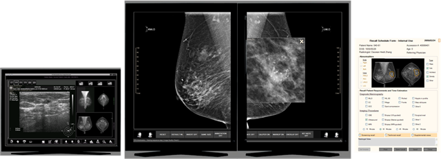
Three Palm Software’s breast imaging workstation product¹ is called WorkstationOne, and offers a distinctly different style of enterprise integration and interpretation workflow. Three Palm Software understands the data-intensive challenges that radiologists are facing when moving to digital mammography, and the need to review digital mammograms as efficiently as they can read films on motorized light boxes. This mammography workstation’s elegant and simple interface is designed to meet the most demanding radiologists’ needs.
User advantages:
- “Single-click” workflow* to streamline mammogram reading from opening a study to generating a report.
- Customized mammographic-specific reading and hanging protocol sequences (including multiple prior support).
- Incorporates expert viewing methodology including Tabár’s systematic viewing mask techniques* for searching for subtle radiographic abnormalities.
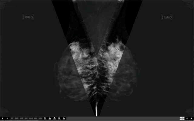
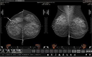
- 8 or more current and prior images are displayed immediately when the next study is opened, with multiple priors accessible by single click.
- An unlimited number of “current” images (2D and 3D) can be stepped through as part of the workflow (e.g., to handle extra views and duplicates), or the user can configure to only see the primary views, with others available with a “page-flip” click.
- Current images are high-lighted in all hanging protocols to minimize the risk of misdiagnosis from priors.
Full-resolution image viewing navigated using a mouse wheel with visual tracing of pixels that have been viewed – no need to manually pan and zoom the images to view all pixels at full resolution:
scrolling to:
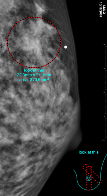
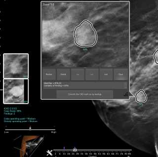
Electronic grease pen allows the radiologist to markup the image display any time, using shapes such as: ellipse; rectangle; free hand; linear measure; arrow; and text. A “markup correlator graphic” can be automatically displayed, allowing the user to visualize the position of a markup drawn in various views – overlaid on an idealized breast outline. Markup can be automatically saved to PACS.
- Range of modalities – digital mammography (all vendors, including Hologic, GE, Siemens, Planmed, Philips, Metaltronica, Medi-Future, Fujifilm, IMS/Giotto), CR (including mammography images from Fuji, Carestream, Agfa, Konica), digitized films, DX, US, MR, CT, NM; integrated CAD, Key Notes and GSPS display.
Tomosynthesis (i.e., 3D) support – tomosynthesis images from most vendors are transferred in DICOM standard format (DBT/BTO), and non-standard Siemens tomosynthesis (CT) is converted automatically for viewing. Breast Projection Images can be included in the workflow. Standard tomo display tools such as cine, manual scrolling, slabbing, along with a 3D navigation icon are supported. Standard 2D mechanisms such as markup (including the correlator), overlay and keynotes are naturally extended to 3D, with the workflow mechanisms automatically including tomo images without requiring user configuration. Enhanced tomo navigation is also supported for sites with tomo-CAD (see below), or with the GE Enhanced “V-Preview” or Hologic “Smart Mapping”.
CAD display is customized for all common systems (more than 15 vendors) including breast density information. Custom graphics and information from each vendor is shown. This support extends to 3D (Tomosynthesis) for vendors that support it. In particular, 3D navigation and display of markers from tomo-CAD is supported for reports from iCAD, and ScreenPoint Medical (with decision support for both 2D and 3D). Tomo navigation includes a keyboard shortcut, thumbnails, shadow-marks, and an assessment popup with more details and navigation.
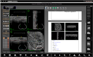
- Collects reporting information automatically as each case is read, with the ability to export that to external systems (RIS, reporting, dictation, etc) or to generate various types of reports internally. For example, the system can generate template-based reports and letters that can be exported in a number of formats including DICOM encapsulated PDF and even email, and automatically generate DICOM structured reports (following the Breast Imaging Template).
- Plug-in viewers for adjunct capabilities such as breast MR CAD, ultrasound CAD, web document review, dictation triggers, view DICOM structured reports, etc.
Plug-in comparison viewer allows display of any mix of modalities and studies in different windows, each with its own tiling, allowing arbitrary comparison (including synchronized scrolling of different studies and modalities).
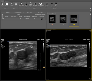
- Optional plug-in viewer for automated breast ultrasound from GE (ABUS), and Siemens (ABVS). This plug-in viewer provides 3D manipulation, markers, and ultrasound report generation.
- Optional plug-in viewer for automated breast ultrasound from iVu (plugin from ORS).
- Mammography film printing – including 10- and 12-bit grayscale, true size, chest-wall alignment, annotation positioning, as required to meet the MQSA guidelines. Multi-image print layouts are also supported.
- Efficient reading due to bi-directional reporting system integration – reading can be driven by an external worklist, and markup and assessment results from the reading are transparently propagated back into the reporting system where they are included in the generated report.
Enterprise features:
- Follows industry standards, compatible with IHE Mammo guidelines.
- Images and related information (such as mammography CAD structured reports and markup defined in GSPS objects) can be pushed to the workstation or can be retrieved from PACS.
- Two levels of pre-fetch – optimistic storage to local disk based on an expected worklist, plus caching to memory during the reading cycle using the interpretation worklist.
- Even with no user logged in, the system automatically pre-fetches studies.
- Multi-reader (local and/or remote) support with automatic and manual arbitration.
- Plug-in mechanism integrates the display of reports from an external RIS and supports the trigger of an external dictation system with the identity of the study being read.
- Peer-to-peer worklist synchronization without the need for a server (reading states on multiple workstations can be automatically synchronized).
IT features:
- Software-only install, so can easily be deployed in the field.
- Runs on a range of Windows platforms (Windows 7 and later) – 64-bit preferred.
- Low maintenance – system pre-fetches and cleans up data automatically.
- Stand-alone configuration tool (allowing service configuration of site settings).
- Intuitive integrated user configuration UI for user options.
- Application honors the security settings of the enterprise – so can work within the policies that an enterprise implements as part of its HIPAA compliance.
- Can work in a mixed environment with workstations from other vendors – e.g., as a “best of breed” viewer to accompany a PACS or VNA.
- Easy user migration from existing workstations (such as SecurView, IDI MammoWorkstation, SenoIris, etc) as WorkstationOne provides even more functionality in an easier to use environment.
OEM vender features:
- Customization capabilities include APIs that expose the ability to provide alternate mechanisms for triggering pre-fetch, work-list integration, and report export.
- No limits on numbers of connections.
Technical data:
- Runs on Windows (7 and later) with the dotnet framework.
- Native 32- and 64-bit versions of the application.
- Recommended to be used with one color monitor for user navigation, and two high-resolution monitors (typically 5MP) for primary diagnosis of mammograms.
- Displays greater than 256 grey levels on suitable monitors, with optimized display on specific display cards which off-load all display tasks from the CPU.
- Includes support for a number of low-level display techniques (to get best speed and quality on the available hardware) – including OpenGL (configurable use of NVIDIA extensions), Direct3D (version 11), ISD (for specific JVC Kenwood monitors), as well as base GDI display mechanisms.
- Acts as a storage SCP in order to receive MG for-presentation studies, Mammography SR reports, GSPS (grayscale softcopy presentation state) objects, and related images in classes such as SC (secondary capture), US (Ultrasound), MR (Magnetic Resonance), NM (Nuclear medicine), CT, DX and the Breast Tomosynthesis IOD (DBT/BTO). Accepts lossless or lossy compression (lossy for MG and DBT – not FDA cleared in the US).
- Acts as storage SCU to output GSPS for radiologist markup, KOS for image correction or teaching purposes, e-PDF, SC and SR for mammography reports or patient letters; negotiates a variety compression schema for image export.
- Acts as a print SCU for film printing to DICOM printers, exporting to DICOM storage or to Windows printers, including customized annotation, overlay, true-size or any layout formatting, grayscale and color output.
- DICOM secure communications (TLS) supported as a configuration option.
- Configurable use of modality worklist, HL7 connections and other mechanisms as hints for upcoming studies (to trigger pre-fetch) and for composing an interpretation worklist.
- Bi-directional HL7 support.
- Launching of cases via http, tcp, and file mechanisms, to support integration with external systems.
- Integration support includes bi-directional worklist synchronization, and export of reporting information.
Notes:
- WorkstationOne has FDA 510K clearance (K073272), CE marking, and other clearances that allow it to be sold in various parts of the world. Contact Three Palm Software for availability in specific regions.
* WorkstationOne features are covered by one or more of TPS’s patents: 8,184,890 8,340,387 8,803,911.